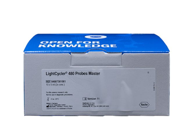
LightCycler® 480 Probes Master
LightCycler® 480 Probes Master is designed for research studies on the LightCycler® 480 System. The LightCycler® 480 Probes Master is a ready-to-use hot start reaction mix designed specifically for detecting DNA targets with hydrolysis probes during LightCycler® 480 System PCR. However, it may be used in other types of PCR on the LightCycler® 480 System. For best results, use this master mix with LightCycler® 480 Multiwell Plates. The kit can also help prevent carryover contamination during PCR (when used with LightCycler® Uracil-DNA Glycosylase*) or to perform the second step of a two-step RT-PCR.
Benefits of the LightCycler® 480 Probes Master
- Perform sensitive, specific, and quantitative PCR or RT-PCR using the LightCycler® 480 Instrument.
- Eliminate time-consuming MgCl2 titration.
- Establish new qPCR assays and get results quickly by combining LightCycler® 480 Probes Master with Universal ProbeLibrary probes for mono- or dual-color quantification assays.
- Save time with a convenient, ready-to-use 2x concentrated hot start master mix.
- Minimize pipetting steps and contamination risk.
How the LightCycler® 480 Probes Master works
LightCycler® 480 Probes Master is a ready-to-use reaction mix specifically developed for the hydrolysis probe detection format in multiwell plates on the LightCycler® 480 Instrument. It contains FastStart Taq DNA Polymerase for hot start PCR, which significantly improves the specificity and sensitivity of PCR by minimizing the formation of nonspecific amplification products (Chou, Q. et al., 1992, Kellogg, D.E. et al., 1994, and Birch, D.E., 1996). FastStart Taq DNA Polymerase is a chemically modified form of thermostable recombinant Taq DNA Polymerase that shows no activity up to 75°C. The enzyme is active only at high temperatures, where primers no longer bind nonspecifically. The enzyme is fully activated (by removal of blocking groups) in a single pre-incubation step (95°C, 5 – 10 minutes) before cycling begins. Activation does not require the extra handling steps typical of other hot start techniques.
Sequence-specific detection of PCR products relies on sequence-specific oligonucleotide probes that are coupled to fluorophores. These probes hybridize to their complementary sequence in target PCR products. Probe chemistries that are suitable for use in the LightCycler® 480 Instrument include single-labeled probes, hybridization probes, and hydrolysis probes. Hybridization and hydrolysis probe chemistries use the so-called FRET principle. Fluorescence Resonance Energy Transfer (FRET) is based on the transfer of energy from one fluorophore (the donor or reporter) to another adjacent fluorophore (the acceptor or quencher).
Hydrolysis probe assays can technically be described as homogeneous 5′-nuclease assays, since a single 3′-non-extendable probe, which is cleaved during PCR amplification, is used to detect the accumulation of a specific target DNA sequence (Holland, P. M. et al., 1991). This single probe contains two labels, a fluorescent reporter and a quencher, in close proximity to each other. When the probe is intact, the quencher dye is close enough to the reporter dye to suppress the reporter fluorescent signal (fluorescent quenching takes place via FRET). During PCR, the 5′-nuclease activity of the polymerase cleaves the hydrolysis probe, separating the reporter and quencher. The reporter dye is no longer quenched and emits a fluorescent signal when excited. The LightCycler® 480 Instrument can detect hydrolysis probes that are labeled with the reporter dyes LightCycler® Red 610, LightCycler® Red 640, LightCycler® Cyan 500, FAM, or HEX. These labeled hydrolysis probes can be used separately or in combination, which permits either single- or multicolor detection.
For multicolor hydrolysis probe assays, it is recommended to use dark quencher dyes (i.e., dye molecules which efficiently quench the fluorescence of a FRET reporter dye without emitting fluorescence themselves). Roche Life Science recommends using BHQ-2 (quenching range 550 -650 nm) for all hydrolysis probe reporter dyes listed above.

Figure: Amplification curves were obtained from serial dilutions (triplicates) of 1 (far right), 10, 50 100, 1,000, 5,000, 10,000, and 100,000 (far left) copies Cytochrome P450 2C9 transcript per well. A specific set of primers and a FAM/TAMRA-labeled hydrolysis probe that recognizes a 196-bp fragment of the Cytochrome P450 2C9 gene was used.
